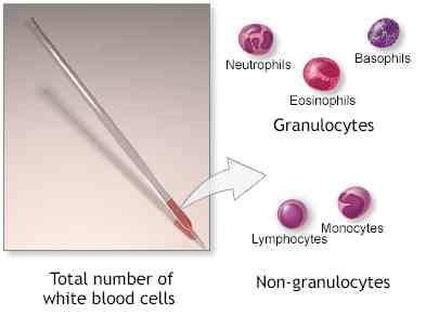
Leucocyte count
- Introduction
2. preparation of blood smear
3. Types of staining
4. staining of blood smear
5. microscopic examination
Introduction of differential leucocyte count.
Differential leucocyte count is important for the diagnosis of various blood related disorder involving white blood cell and red blood cells.
generally, it is performed to check the normal distribution of different leucocyte eg :- neutrophils, basophils, eosinophils, monocyte and lymphocyte .
The blood specimen to be used for differential leucocyte count is done by capillary or EDTA blood.
2. preparation of blood smear
First of all, take a clean glass slide then one drop OF Capillary blood or EDTA is obtained on a corner of the slides.
Take the Spreader with a sharp edge and place it on a drop of the slide at a 45° angle then the spreader is pushed backward so that the Drop is spread evenly to the edge of the Spreader.
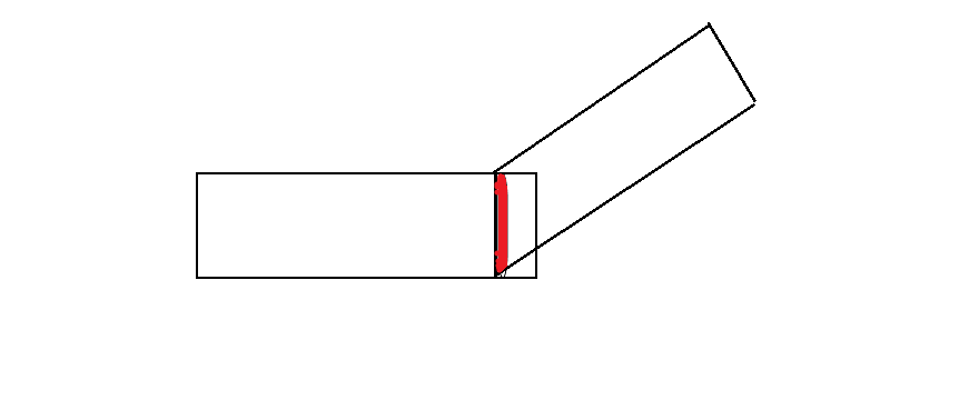
Spread on the slide should be about 45 degrees with a quick movement towards the other end of the slide then the Smear is prepared.
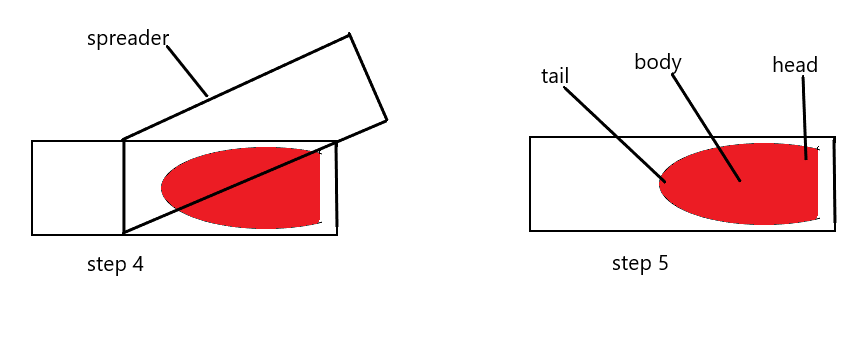
Then perfect smear shape is tongue shape, and is divided into three part head tail and body and and only we are count body part.
3. Types of stain
Leishman stain
Giemsa stain
Wright stain
NOTE:- Leishman stain is commonly used in the laboratory and it is the cheapest of all stain.
Composition:-
Leishman stain powder- 0.15 gm
Methyl alcohol-100 gm
4. Staining of prepared smear:-
After making of blood smear slide it should be dry in the air.
And then it is put on the staining rack.
Then smear is covered with Leishman stain and waited for 2 minutes.
After 2 minutes add distilled water or buffer solution and mix with stain on the smear slide and wait for 8 to 10 minutes.
After 10 minute wash the slide in the distilled water to remove Excess stain.
Then air dry the making film and observe under the microscope.
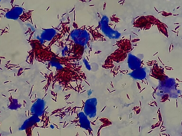
5. Microscopic Examination:-
First, examine the stained blood Smear under low power objective Lens.
Staining smear 3 Jones can be identified.
a. Identified area head of smear
b. The central area is the body.
c. The end of the smear is a tail region.
Choose the portion of the smear in the body region slightly Before the tail end.
Observe the slides under the oil emersion objective lens by putting a drop of oil over the slides.
Count each type of white cell observed and record the observation on a piece of paper in a tabular form and continue till the 100 cells are counted this will give the average number of white blood Cells.
6. Observation of differential leucocyte count:-
- Neutrophils: – purple color nuclei with pink color cytoplasm.
- Eosinophil:- cytoplasm is a faint pink color.
- Basophil:- granules stain dark blue with purple nuclei.
- Monocyte:- pink cytoplasm with purple color nucleus.
- Lymphocyte:- dark blue nucleus with light blue cytoplasm.
- Platelets:- Violet color Granules.
- Red blood cell:- pink color.
leucocyte
This is colorless.
Nucleated cell.
The number of WBCs in blood is 4000 to 11000.
Two types
Granulocyte
A granulocyte
Granulocyte:-
Based on the shape of the nucleus and die different types.
Neutrophils
Basophils
Eosinophils
Neutrophils:-
Neutrophils or polymorph:-
60 to 70% of the total WBC.
Diameter 10 to 12 microns
Number of lobes 2 to 7.
Life span- 3 to 5 days.
Function:- phagocytic.
Fine granules.
Basophils:-
Normal range 0 to 1 %.
Life span is 12 to 15 days.
Diameter 8 to 10 microns.
The nucleus is not clear.
Bilobed kidney shaped.
Eosinophils:-
Size 12 to 15 microns in diameter.
Shape rounded.
Lobe -2
Granule – dense
Normal range 1 to 5 %
Life span 7 to 12 days.
Agranulocyte:-
Monocyte
Lymphocyte
Monocyte:-
Largest cell among 5, the shape is irregular or is not well founded.
Size 14 to 18 microns in diameter.
Range 2 to 8%
Shape kidney or notch shape.
Lymphocyte:-
T Lymphocyte: –
The nucleus is large and occupies 90% of the total cell area.
Size 7 to 10 micron in Diameter.
Shape rounded.
B Lymphocyte: –
Nucleus Lie to one side of the cell.
Shape rounded.
Size 10 to 12 microns in diameter.
Range 25 to 35%.
what is the normal leucocyte count?
normal leucocyte count is 4000 to 11000/cu mm
what is differential leucocyte count is high?
it means you have an infection, and you can suffer from disease and any allergic reaction.
If WBC count is to high ?
in one micro liter blood contain more than 11000 thousand WBC count then its consider leuco-cytosis.