
Microscopic examination of urine
It is studied in the following steps: –
1. Introduction
2. Principle
3. Requirement
4. Instrument
5. Specimen
6. Procedure
7. Observation under a microscope
Introduction of Microscopic examination of urine:-
The microscopic examination is a valuable tool for the detection and evaluation of renal and urinary tract disorders and another systematic disorders.
The average volume of urine is about 1200 ml per day.
Principle: –
The microscopic elements present in urine are collected in the form of deposits by centrifugation a small drop of the sediment is examined by making a weight Preparation of urine and observing under the microscope.
Requirement: –
Test tube
Glass slide
Coverslip
Pasture pipette
Micropipette
Urine container
Instrument: –
Microscope
Centrifuge machine
Specimen:-
collection of urine in the first morning time some urine is avoided and then collected in a sterilized container.
Preservation: –
It should be stored at 2° to 8° degrees Celsius in a refrigerator.
Procedure: –
- Take urine in a clean and dry test until 5 ml.
- Centrifuge for 2500 RPM at 5 minutes after the balanced test tube.
- The supernatant liquid is discarded in another test tube for preparation of protein and deposit material take one drop with the help of a Pasture pipette ya micropipette on a glass slide.
- And then a specimen of One drop is covered with a cover slip.
- It was observed first under a low-power objective lens for the fix.
- After fixing the nose piece for a 40x high power objective lens and saw the following types of sediment organized and unorganized.
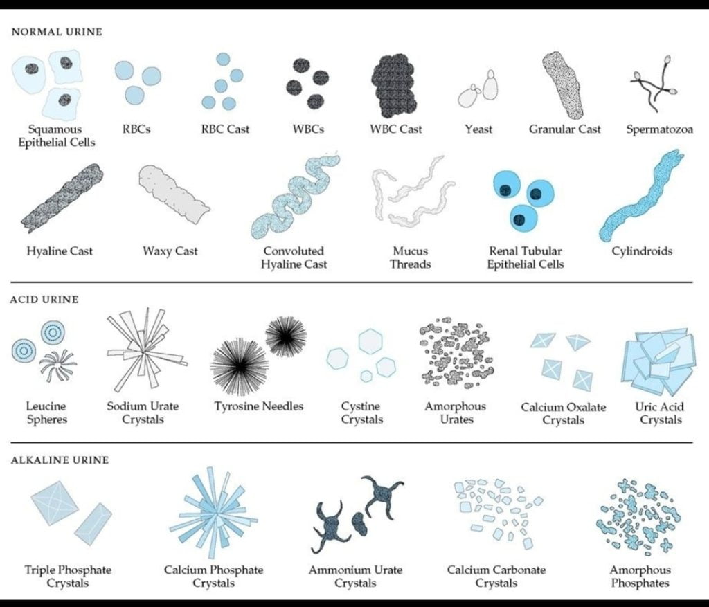
Organized:-
- Pus cell
- Erythrocyte
- Cast
- Epithelial cell
- Bacteria, parasites, fungi
Unorganised:-
- Crystals
- Amorphous
Observation:-
1. Pus cell (wBC):-
It is also known as a leucocyte.
Normal pus cells contain urine 2 to 3 cells/per hot ( high power field).
These are mostly neutrophils.
2. Erythrocyte:-
Impress urine the colour of sales normal pale or yellow appearance.
they appear smooth by a concave disk about a cell micrometer in diameter.
They do not contain nuclei.
Erythrocytes are not usually present in normal disease.
3. Cast:-
These are formed by the coagulation of albuminous material and the renal tubules.
The cast is cylindrical and varies in diameter.
Cast have nearly parallel sides and rounded or blunted ends.
Cast dissolves in alkaline urine.
They may be convoluted straight or curves.
There are different types of cast
Hyaline cast
Red cell
Granular
Epithelial
Waxy
Fatty
4. Crystal’s:-
Crystal found in Abnormal urine and metabolic disorder.
Normal Crystal’s: –
Uric acid
Calcium oxalate
Hippuric acid
Calcium phosphate
Triple phosphate
Calcium carbonate
Ammonium biuret
Calcium sulphate
Abnormal Crystal:-
Bilirubin
Cholesterol
Cystine Crystal
Leucine
Types in
Microscopic examination of urine ,Microscopic examination of urine,Microscopic examination of urine